
Mitral valve repair is surgery that restores the function of a patient’s own mitral valve rather than removing it and fitting an artificial one. When the valve anatomy is suitable and a durable repair can be expected, preserving the native valve is generally preferred to replacement.
This guide explains how the mitral valve works, the conditions that damage it, how surgeons decide when to operate, the repair techniques used today, and what recovery and long-term follow-up involve.
Medical note: This article is patient education and is not a substitute for individual medical advice. Whether a valve can be repaired — and when surgery should be performed — can only be decided after a full assessment of the valve, the heart and the patient’s overall risk.
What Is Mitral Valve Repair?
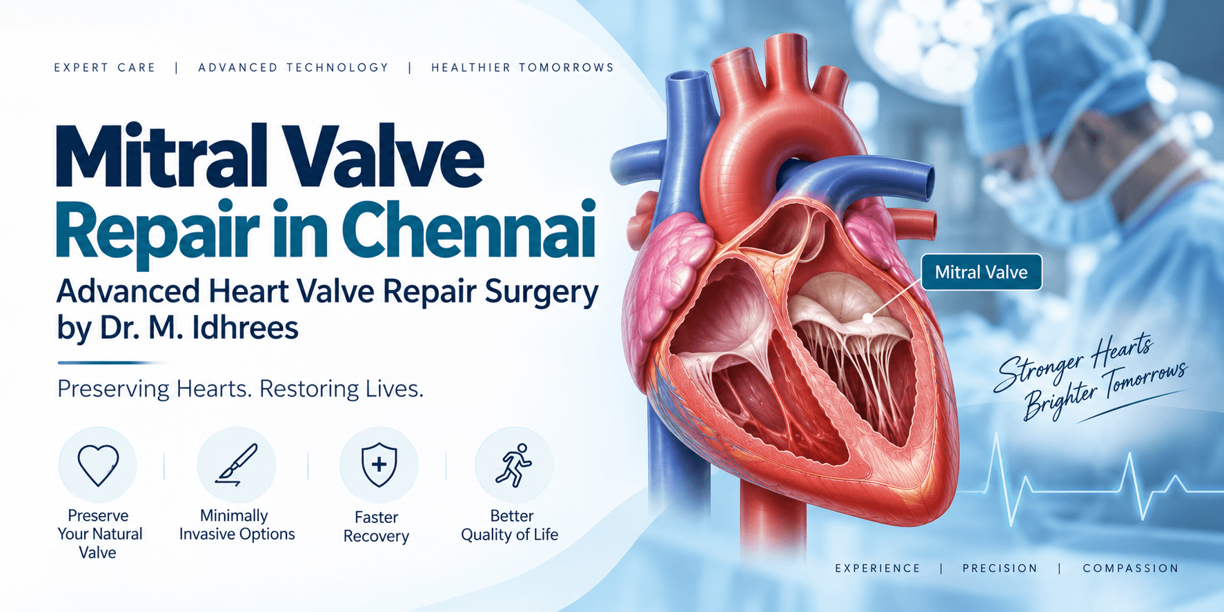
The mitral valve sits between the left atrium and the left ventricle. Oxygen-rich blood returns from the lungs into the left atrium, passes through the mitral valve into the left ventricle, and is then pumped out through the aortic valve to the rest of the body.
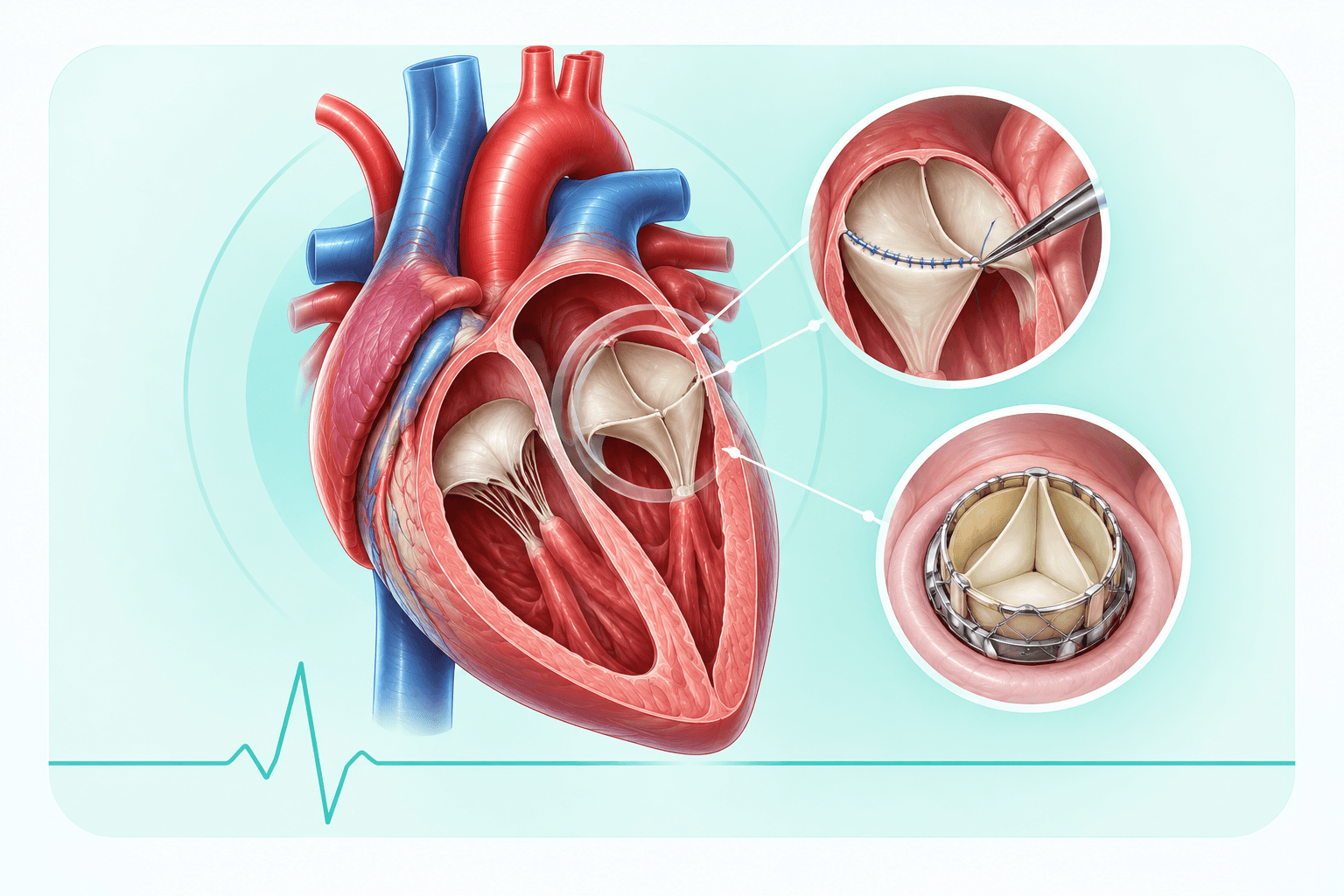
The valve has two leaflets, supported by a ring of tissue called the annulus and by fine cords — the chordae tendineae — that anchor the leaflets to the papillary muscles inside the ventricle. Each part has to work together for the valve to open freely and then close completely with every heartbeat.
When any of these structures is damaged, the valve may leak backward (regurgitation), fail to open fully (stenosis), or both. Mitral valve repair reconstructs the damaged parts — the leaflets, the chordae or the annulus — so that the patient’s own valve closes properly again.
Why Preserving the Native Valve Matters
The mitral valve is not simply a door. Its cords and attachments are part of how the left ventricle contracts and holds its shape. Preserving these structures helps maintain ventricular function after surgery.
A repaired valve also avoids the trade-offs that come with a prosthesis. Mechanical valves usually require lifelong anticoagulation; tissue valves avoid that in most patients but have a limited lifespan and may eventually need reintervention. Where a durable repair is achievable, it avoids both.
For these reasons, current practice favours repair over replacement in suitable patients — particularly in degenerative mitral regurgitation — when an experienced team expects a durable result.
Understanding Mitral Valve Disease
Mitral Regurgitation
Mitral regurgitation means the valve leaks, allowing blood to flow back from the left ventricle into the left atrium with each heartbeat. The heart compensates by pumping a larger volume, and over time the left atrium and left ventricle enlarge. Symptoms typically develop gradually — breathlessness on exertion, fatigue, palpitations — and may appear late, after the heart has already begun to change.
Regurgitation is described as primary (degenerative) when the valve itself is diseased, and secondary (functional) when the valve leaks because the ventricle or annulus has enlarged. The distinction matters, because it changes both the treatment options and the timing of surgery.
Mitral Valve Prolapse
In mitral valve prolapse, one or both leaflets bulge back into the left atrium when the valve should be closed, often because the tissue is redundant or the supporting chordae are elongated. Many people with prolapse have little or no leakage and need only periodic review. Others develop progressive regurgitation, and prolapse is one of the most repairable forms of mitral valve disease.
Mitral Stenosis
Mitral stenosis means the valve does not open widely enough, restricting flow from the left atrium into the ventricle. Pressure builds back into the lungs, causing breathlessness, reduced exercise capacity and sometimes coughing or episodes of blood-streaked sputum. Atrial fibrillation is common as the left atrium enlarges. Rheumatic heart disease remains the most frequent cause worldwide.
Infective Endocarditis
Infection of the valve can destroy leaflet tissue, rupture chordae or create abscesses and perforations, producing severe regurgitation over days or weeks rather than years. Treatment combines prolonged antibiotics with surgery when the valve is destroyed, the infection is uncontrolled, or the patient develops heart failure or embolic complications.
Degenerative Mitral Valve Disease
Degenerative disease covers changes in the valve tissue itself — thickened or redundant leaflets, stretched or ruptured chordae, and calcification of the annulus. It is the most common cause of primary mitral regurgitation in many populations, and the group in which repair is most often possible.
Rheumatic Mitral Valve Disease
Rheumatic fever after streptococcal infection can scar and fuse the leaflets and cords over many years, producing stenosis, regurgitation or a mixed lesion. Rheumatic valves are more difficult to repair than degenerative ones because the tissue itself is scarred, but repair remains possible in selected patients, particularly younger ones.
Signs and Symptoms
Mitral valve disease often progresses quietly. Many patients adjust their activity without realising it, which is why symptoms are best assessed against what a person could comfortably do a year or two earlier.
Early Symptoms
- Fatigue and reduced stamina
- Mild breathlessness on exertion
- Palpitations or an irregular heartbeat
Progressive Symptoms
- Chest discomfort
- Swelling of the feet and ankles
- Reduced exercise tolerance
- A persistent cough, particularly when lying flat
- Waking at night short of breath
Symptoms Needing Emergency Assessment
- Severe or sudden breathlessness at rest
- Pulmonary oedema — frothy sputum and severe difficulty breathing
- Fainting or collapse
Sudden severe symptoms can indicate acute mitral regurgitation, for example after chordal rupture or in endocarditis, and need immediate hospital assessment.
Who Needs Mitral Valve Repair?
Surgery is considered when the leak or obstruction is severe and the heart is affected, or is likely to become affected if the condition continues. Assessment usually weighs:
- Severe mitral regurgitation with symptoms
- Enlargement of the left ventricle or left atrium
- A falling left ventricular ejection fraction
- Symptomatic prolapse with a flail or severely prolapsing leaflet
- Chordal rupture causing acute severe regurgitation
- Infective endocarditis with valve destruction, heart failure or uncontrolled infection
- New atrial fibrillation or rising pulmonary artery pressure
- The likelihood that the valve can be repaired durably
- The patient’s age, other medical conditions and overall surgical risk
In asymptomatic severe regurgitation, the case for early surgery is strongest when a durable repair is expected by an experienced team, because operating before the ventricle deteriorates protects long-term heart function.
Diagnosis
Echocardiography
A transthoracic echocardiogram is the first and most important test. It shows the valve’s structure and movement, grades the severity of regurgitation or stenosis, and measures left ventricular size and function, left atrial size and pulmonary pressures.
Transoesophageal Echocardiography (TEE)
A TEE gives a far more detailed view of the leaflets, the chordae and the annulus, including three-dimensional images of the valve. It is central to surgical planning — it identifies exactly which segment of which leaflet is diseased — and is also used in the operating theatre to confirm the result of the repair before the chest is closed.
CT Scan
CT assesses annular calcification, chest wall and rib anatomy, and the aorta and peripheral vessels. This matters particularly when minimally invasive or robotic access is being considered.
Cardiac MRI
MRI can quantify regurgitant volume and measure ventricular volumes and function accurately when echocardiographic findings are unclear or inconsistent with the clinical picture.
Coronary Angiography and Cardiac Catheterisation
Coronary angiography is performed before surgery in patients at risk of coronary disease, so that any bypass grafting can be planned in the same operation. Catheterisation can also measure pressures directly when non-invasive findings are inconclusive.
Blood Tests and Other Investigations
Blood tests assess kidney, liver and thyroid function, anaemia and clotting, and blood cultures are essential when endocarditis is suspected. An ECG identifies atrial fibrillation and other rhythm problems, and dental assessment is usually arranged before valve surgery.
Advanced Mitral Valve Repair Techniques
Mitral repair is not a single operation. The surgeon selects and combines techniques according to which part of the valve has failed, and confirms the result with echocardiography before leaving the operating theatre.
Annuloplasty Ring Repair
A prosthetic ring or band is sewn around the mitral annulus to restore its normal size and shape so the leaflets meet properly. Annuloplasty is used in most mitral repairs, either on its own in functional regurgitation or alongside leaflet and chordal work, and it reduces stress on the rest of the repair.
Chordal Replacement with Artificial Chordae
Elongated or ruptured cords are replaced with expanded PTFE (Gore-Tex) sutures anchored to the papillary muscle and the leaflet edge. Setting the correct length restores the leaflet to its normal position. This technique preserves leaflet tissue and is widely used in prolapse and flail leaflet repair.
Chordal Transfer
A healthy secondary cord, or a cord from an adjacent segment, is transferred to support the prolapsing part of the leaflet. It is an alternative to artificial chordae using the patient’s own tissue.
Triangular Resection
A small triangle of prolapsing leaflet tissue is removed and the edges are sewn together. It is a tissue-sparing technique suited to localised prolapse, most often of the anterior leaflet or a limited posterior segment.
Quadrangular Resection
A rectangular segment of a prolapsing posterior leaflet is removed and the leaflet is reconstructed, usually with a sliding plasty to redistribute the remaining tissue and reduce the risk of systolic anterior motion. It is a long-established technique for posterior leaflet prolapse.
Leaflet Reconstruction
Perforations or tissue defects — commonly after endocarditis — are repaired with a patch of the patient’s own pericardium, restoring a competent closing surface.
Commissurotomy
In stenosis, fused commissures are divided and thickened cords are split so the valve opens more widely. This is most often used in rheumatic disease, sometimes combined with decalcification and annuloplasty.
Calcium Removal (Decalcification)
Calcium deposits in the annulus or leaflets are carefully removed to restore leaflet mobility. Extensive annular calcification makes repair more demanding and is one of the factors that can favour replacement.
Edge-to-Edge Repair
The free edges of the two leaflets are stitched together at the point of the leak, creating a double-orifice valve. It is used in selected anatomies, often as an adjunct to annuloplasty. The same principle underlies transcatheter edge-to-edge devices used in patients who are not candidates for surgery.
Minimally Invasive Mitral Valve Repair
The valve is repaired through a small right-sided incision between the ribs rather than a full sternotomy, using specialised instruments and camera assistance. In suitable patients this means less surgical trauma, reduced blood loss, a smaller scar and a shorter hospital stay, with the same repair techniques performed inside the heart.
Robotic Mitral Valve Repair
Robotic assistance adds magnified three-dimensional vision and instruments with a wide range of movement through very small incisions. Candidate selection is more restrictive — chest anatomy, peripheral vessel quality, lung function and valve pathology all matter — and the approach requires a team with specific training and experience.
Mitral Valve Repair vs Replacement
| Feature | Repair | Replacement |
|---|---|---|
| Native valve preserved | Yes | No |
| Effect on heart function | Subvalvular structures preserved, supporting ventricular function | Function preserved when the subvalvular apparatus is retained |
| Lifelong blood thinners | Usually not required long term | Required for mechanical valves; usually not for tissue valves |
| Risk of prosthetic infection | No prosthetic leaflets implanted | Prosthetic valve endocarditis is a recognised risk |
| Durability | Good long-term durability in suitable anatomy, most established in degenerative disease | Mechanical valves are durable; tissue valves degenerate over time |
| Best suited to | Degenerative disease, prolapse, chordal rupture, selected rheumatic and infective cases | Heavily calcified, scarred or destroyed valves, and failed or unsuitable repairs |
Replacement is the right operation in many patients, and choosing it is not a lesser outcome — a valve that cannot be repaired durably is better replaced than repaired badly. The decision is made from the imaging, the findings at surgery and the surgeon’s assessment of what will last.
Why Repair Is Generally Preferred
- The valve’s own tissue and supporting cords are preserved, which helps maintain left ventricular function
- Most patients avoid lifelong anticoagulation and its bleeding risk
- No prosthetic leaflets are implanted, avoiding prosthetic valve degeneration and prosthetic endocarditis
- Long-term outcomes after successful repair for degenerative disease compare favourably with replacement
- Anticoagulation-free recovery is particularly valuable in younger and physically active patients
These advantages depend on the repair being durable. That is why the likelihood of a successful repair — which depends on both the valve anatomy and the team’s experience — is part of the decision about where and when to operate.
What Happens During Surgery?
Anaesthesia and Monitoring
The operation is performed under general anaesthesia. Monitoring lines are placed and a transoesophageal echocardiography probe is positioned to assess the valve before and after the repair.
Surgical Access
Access is either through a sternotomy or, in selected patients, through a small right-sided incision between the ribs for a minimally invasive or robotic approach.
Cardiopulmonary Bypass
A heart-lung machine takes over the work of the heart and lungs so that the heart can be stopped and opened. The heart is protected with a cardioplegia solution during this time.
The Repair
The left atrium is opened and the valve is inspected directly. The surgeon identifies the failed segment and performs the appropriate combination of leaflet, chordal and annular work, usually completed with an annuloplasty ring.
Intraoperative Echocardiography
Once the heart is beating again, TEE is used to confirm that the valve closes properly, that any residual leak is minimal and that there is no obstruction. If the result is not satisfactory, the surgeon can revise the repair or proceed to replacement during the same operation.
Closure and ICU Transfer
Drains and temporary pacing wires are placed, the chest is closed, and the patient is transferred to the intensive care unit, usually still on a ventilator for a short period while waking from anaesthesia.
Recovery Timeline
Recovery varies with age, heart function before surgery, the surgical approach and other medical conditions. The following is a general guide rather than a fixed schedule.
- Days 1–2: Intensive care. Breathing tube and drains removed as soon as it is safe; sitting out of bed and early breathing exercises begin
- Days 3–7: Ward recovery. Walking short distances, chest physiotherapy, adjustment of medications and rhythm monitoring before discharge
- Week 2: A structured walking programme at home, with distance increased gradually
- Week 4: Light daily activities resumed; driving usually discussed at the follow-up review
- Weeks 6–8: Many patients return to office work; heavy lifting is still avoided while the sternum heals
- 3 months: Most patients reach near-complete recovery, with exercise capacity continuing to improve
Recovery after a minimally invasive or robotic repair is often quicker, because there is no sternal healing to protect.
Risks and Complications
Mitral valve repair is a major cardiac operation, and the risks are real but generally low in appropriately selected patients operated on by an experienced team.
- Bleeding, occasionally requiring transfusion or a return to theatre
- Atrial fibrillation and other rhythm disturbances, which are common after valve surgery and often temporary
- Stroke or other neurological complications
- Wound, chest or bloodstream infection
- Residual or recurrent valve leak
- Need for valve replacement during the same operation if the repair is not satisfactory
- Reoperation at a later date, which is uncommon after a durable repair
- Kidney, lung or other organ complications, more likely in patients with existing disease
Careful assessment before surgery, detailed imaging, appropriate timing, intraoperative echocardiographic checking of the repair and structured postoperative care all reduce these risks. Operating before the ventricle has deteriorated is one of the most effective ways to improve the outcome.
Long-Term Results
A successful mitral repair is durable in most patients, and the majority never need further valve surgery. Recurrent regurgitation is possible, particularly where the underlying disease continues — for example in functional regurgitation from an enlarged ventricle, or in rheumatic disease.
- Follow-up: a clinical review and echocardiogram before discharge, again in the early months after surgery, and then at intervals advised by the team — usually yearly once the result is stable
- Medication: rhythm and blood pressure medications as prescribed; anticoagulation for a limited period after some repairs, and longer where atrial fibrillation is present
- Exercise: a graded return to activity, ideally through cardiac rehabilitation, building to regular aerobic exercise
- Lifestyle: blood pressure control, stopping smoking, a heart-healthy diet, weight management and treatment of diabetes
- Endocarditis prevention: good dental hygiene, regular dental review, and prompt treatment of infections. Ask the treating team whether antibiotic prophylaxis before dental procedures applies in your case
Report any new breathlessness, palpitations, swelling, fever or reduced exercise tolerance to the treating team rather than waiting for the next scheduled review.
Why Choose Dr. M. Idhrees
Dr. Mohammad Idhrees is a cardiac and aortic surgeon whose practice includes heart valve repair and replacement alongside complex aortic surgery. Patients being assessed for mitral valve disease receive:
- Detailed valve assessment with echocardiography and, where needed, TEE, CT and MRI before any decision on surgery
- A clear discussion of whether the valve is likely to be repairable, and of repair versus replacement in their specific case
- Experience with complex valve reconstruction and with minimally invasive cardiac surgery in suitable patients
- A focus on preserving the patient’s own valve wherever a durable repair can be expected
- Multidisciplinary planning with cardiology, anaesthesia and intensive care, and individualised follow-up after surgery
Frequently Asked Questions
- What is mitral valve repair?
- It is heart surgery that reconstructs a patient’s own mitral valve — the leaflets, the supporting cords and the annulus — so that it opens and closes normally again, instead of removing it and fitting an artificial valve.
- Is repair better than replacement?
- When a durable repair is achievable, it is generally preferred, because it preserves the native valve and its supporting structures and usually avoids lifelong anticoagulation. When the valve is heavily calcified, scarred or destroyed, replacement is the better operation.
- How do I know whether my valve can be repaired?
- Echocardiography, particularly transoesophageal echocardiography, shows which part of the valve has failed and how much healthy tissue remains. That assessment, together with the surgeon’s experience, determines how likely a durable repair is. The final confirmation comes when the valve is inspected during surgery.
- How long does mitral valve repair surgery take?
- Most operations take several hours, including anaesthesia, the repair itself and the echocardiographic checks afterwards. The exact time depends on the complexity of the valve disease, the surgical approach and whether other procedures are performed at the same time.
- How painful is the recovery?
- Discomfort is expected in the first days and is managed with prescribed pain relief. Most patients describe it as manageable soreness rather than severe pain, and it improves steadily over the following weeks.
- How long will I stay in hospital?
- A typical stay is around a week, starting with a short period in intensive care. It may be shorter after a minimally invasive repair, or longer if complications such as atrial fibrillation need treatment.
- Will I need blood thinners after mitral valve repair?
- Many patients take anticoagulation for a limited period after repair, and some do not need it at all. Long-term anticoagulation is usually needed only when there is another reason for it, such as atrial fibrillation. This is decided by the treating team.
- Can mitral regurgitation come back after repair?
- It can, though it is uncommon after a successful repair for degenerative disease. Recurrence is more likely when the underlying cause continues, such as functional regurgitation from an enlarged ventricle or ongoing rheumatic disease. Regular echocardiographic follow-up detects it early.
- Is robotic mitral valve repair better?
- Robotic and minimally invasive approaches avoid a sternotomy and can mean less pain, a smaller scar and quicker recovery. They are not suitable for every patient or every valve, and the quality and durability of the repair inside the heart matters more than the size of the incision.
- Am I too old for mitral valve repair?
- Age alone does not decide suitability. Overall health, heart and lung function, kidney function and frailty matter more, and many older patients do well after valve surgery. Where surgical risk is high, transcatheter options may be considered.
- What happens if I don’t have surgery?
- Severe mitral valve disease usually progresses. Continued volume or pressure overload can enlarge and weaken the left ventricle, raise pulmonary pressures and lead to atrial fibrillation and heart failure. Some of that damage does not fully reverse once it has occurred, which is why timing matters.
- Can I live a normal life after mitral valve repair?
- Most patients return to their normal activities, including work, travel and exercise, once recovery is complete. Long-term follow-up and attention to blood pressure, rhythm and dental health remain important.
- When can I go back to work?
- Many people doing office-based work return around six to eight weeks after surgery. Physically demanding work usually takes longer, and the treating team will advise based on the approach used and your recovery.
- When can I drive again?
- Driving is usually discussed at the first follow-up review, commonly around four weeks after surgery, once you are off strong pain relief, can move comfortably and could perform an emergency stop.
- Can I exercise after mitral valve repair?
- Yes, and regular exercise is encouraged once you have recovered. Start with the walking programme given at discharge, use cardiac rehabilitation where it is available, and avoid heavy lifting until the sternum has healed if you had a sternotomy.
- When can I travel or fly after surgery?
- Short journeys are usually possible within a few weeks. Air travel is commonly deferred until the early recovery period is complete and the follow-up review has taken place. Ask the treating team before booking, particularly for long flights.
- What should I eat after mitral valve surgery?
- A heart-healthy diet: plenty of vegetables, fruit, whole grains and legumes, adequate protein for healing, limited salt, and limited processed and fried food. If you are taking warfarin, keep your intake of vitamin K-rich foods consistent rather than avoiding them.
- Will I have a large scar?
- A sternotomy leaves a vertical scar down the centre of the chest, which fades over time. Minimally invasive and robotic approaches leave much smaller scars on the right side of the chest.
- Is atrial fibrillation after surgery normal?
- It is common after cardiac surgery and is often temporary, treated with medication and sometimes a controlled restoration of normal rhythm. Persistent atrial fibrillation may need longer-term rhythm control and anticoagulation.
- Can atrial fibrillation be treated at the same time as valve surgery?
- Yes. In selected patients with existing atrial fibrillation, an ablation procedure such as the maze procedure, and management of the left atrial appendage, can be performed during the same operation.
- How often do I need scans after mitral valve repair?
- An echocardiogram is performed before discharge and again in the months after surgery. Once the result is stable, yearly echocardiography is typical, with more frequent imaging if there is a residual leak or the heart function is being watched closely.
- Do I need antibiotics before dental treatment?
- Recommendations depend on what was implanted and on your individual risk, and differ between guidelines. Good dental hygiene and regular dental review matter in every case. Ask your surgeon or cardiologist what applies to you, and tell your dentist about your valve surgery.
- What is the difference between mitral regurgitation and mitral stenosis?
- Regurgitation means the valve leaks backward when it should be closed. Stenosis means the valve does not open widely enough. Some patients have both.
- Is mitral valve prolapse dangerous?
- Most people with mitral valve prolapse have little or no leakage and remain well with periodic review. It becomes significant when it causes progressive regurgitation, symptoms or changes in heart size and function.
- What is a flail mitral leaflet?
- A flail leaflet occurs when the cords supporting it rupture, so part of the leaflet moves freely into the left atrium. It usually causes severe regurgitation, sometimes suddenly, and is often well suited to repair with artificial chordae.
- Can mitral valve disease be treated with medication alone?
- Medication can control symptoms, heart rate and blood pressure, and treat heart failure, but it cannot fix a structurally damaged valve. In severe disease, medication is supportive rather than a substitute for surgery.
- What is transcatheter mitral repair, and is it an alternative?
- Transcatheter edge-to-edge repair clips the leaflets together through a catheter rather than open surgery. It is used mainly in patients at high surgical risk or with functional regurgitation, and is considered by a heart team rather than as a routine alternative to surgical repair.
- When should I seek urgent medical attention after surgery?
- Contact the treating team or attend emergency care for severe or sudden breathlessness, chest pain, fever, a discharging or opening wound, fainting, sustained palpitations, or rapidly increasing swelling of the legs.
Conclusion
Mitral valve repair preserves a patient’s own valve and the structures that support the left ventricle, and in suitable anatomy it offers durable results without lifelong anticoagulation for most patients.
The two decisions that matter most are whether the valve can be repaired durably and when to operate. Both depend on detailed imaging, careful assessment of heart size and function, and the experience of the surgical team.
If you have been diagnosed with mitral regurgitation, mitral valve prolapse or mitral stenosis, an early specialist assessment will clarify whether continued monitoring, repair or replacement is the right course for you.