Minimally invasive heart surgery is an advanced approach that allows complex heart procedures to be performed through small incisions between the ribs, instead of opening the entire breastbone. Compared with traditional open-heart surgery it may offer reduced pain, smaller scars, a lower infection risk and a faster return to everyday activities in appropriately selected patients.
Dr. Idhrees, Senior Cardiovascular and Thoracic Surgeon with over 18 years of experience, offers advanced minimally invasive cardiac procedures for valve disease, congenital heart defects, selected coronary artery disease and other complex heart conditions, using modern evidence-based surgical techniques.
What it is
What is minimally invasive heart surgery?
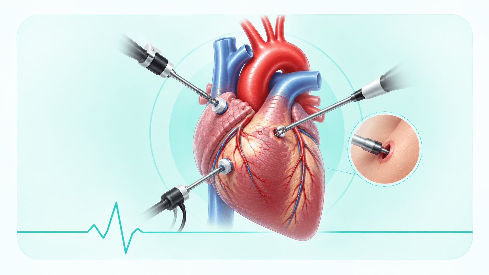
In conventional heart surgery the breastbone is divided down its length to reach the heart. Minimally invasive cardiac surgery reaches the same structures through a small opening — usually between the ribs — leaving the breastbone intact or only partly divided. The operation performed on the heart itself is the same; what changes is the route taken to get there.
Small incisions
Access is typically through an incision of a few centimetres between the ribs, rather than the full-length chest incision of conventional surgery.
No full sternotomy
Because the breastbone is not divided, there is no healing bone to protect afterwards — which is what removes many of the lifting and driving restrictions of the first months.
Specialised instruments
Long, fine, angled instruments designed to work through a narrow opening allow the surgeon to suture and repair at depth without a wide exposure.
Camera-assisted surgery
A high-definition thoracoscope carries a magnified view of the heart to a screen, often showing the valve in more detail than the naked eye would through an open chest.
Robot-assisted surgery
In selected cases, robotic arms controlled by the surgeon translate hand movements into very precise instrument movement inside the chest, with tremor filtered out.
Faster recovery
Less tissue disruption generally means less pain, a shorter hospital stay and an earlier return to work, in patients suitable for the approach.
Conditions treated
Conditions treated with minimally invasive heart surgery
A wide range of procedures can now be performed through a small incision in suitable patients:
Mitral valve repair
Aortic valve replacement
Atrial septal defect (ASD) closure
Patent foramen ovale (PFO) closure
Maze procedure for atrial fibrillation
Removal of cardiac tumours such as atrial myxoma
Selected coronary artery bypass grafting
Tricuspid valve surgery
Mitral valve repair is the procedure where the minimally invasive approach is most established, because the camera view through a right-sided incision shows the valve particularly well.
Types
Types of minimally invasive cardiac surgery
Robot-assisted heart surgery
Instruments on robotic arms are passed through small ports and controlled by the surgeon from a console with a magnified three-dimensional view. Reserved for carefully selected cases where its precision offers a clear advantage.
Thoracoscopic surgery
The operation is guided almost entirely by a camera placed through a port in the chest wall, with instruments through further small openings. It gives the smallest incisions of any approach.
Mini thoracotomy
A small incision between the ribs, most often on the right side of the chest, used for mitral and tricuspid valve surgery and for closing septal defects. The breastbone is left completely intact.
Mini sternotomy
Only the upper part of the breastbone is divided, leaving the lower portion and the chest wall stable. Commonly used for aortic valve surgery, it provides familiar access with far less bone disruption.
Keyhole heart surgery
The general term for these camera-assisted approaches through small openings. Which one is chosen depends on the structure being treated and on your own anatomy.
Benefits
Benefits of minimally invasive heart surgery
The advantages follow from one fact: less healthy tissue is disturbed in order to reach the heart.
A smaller incision
A few centimetres between the ribs rather than the length of the breastbone, which means less trauma to the chest wall and a more stable chest from the first day.
Less blood loss
A smaller surgical field bleeds less, which reduces the likelihood of needing a blood transfusion.
Reduced infection risk
Leaving the breastbone intact removes the possibility of deep sternal wound infection, a complication that is uncommon but serious when it occurs — and a particular concern in patients with diabetes.
Less pain after surgery
There is no healing bone to move with every breath and cough, so most patients need less pain relief and find it easier to breathe deeply and clear their chest.
A shorter stay in intensive care
Patients are often extubated sooner and step down to the ward earlier, because less physical insult means less physiological disturbance to recover from.
Faster overall recovery
Without an eight-to-twelve-week wait for the breastbone to knit, everyday activity, lifting and driving generally resume considerably sooner.
An earlier return to work
Many patients return to desk-based work in a matter of weeks, which matters especially to younger patients and the self-employed.
A better cosmetic result
The scar is small and can often be positioned to sit discreetly along the contour of the chest — not the main reason to choose the approach, but a real benefit for many patients.
Who is suitable
Who is a candidate for minimally invasive heart surgery?
Suitability is decided on your imaging, not on preference. The surgeon has to be satisfied that the structure needing treatment can be reached and repaired just as completely through the smaller opening — the approach is never allowed to compromise the quality of the operation.
Patients who are often suitable include those with:
Mitral or tricuspid valve disease, particularly valves suitable for repair
Aortic valve disease needing replacement, via a mini sternotomy
Congenital defects such as an ASD or PFO
Coronary disease limited to vessels reachable through a small incision
Earlier-stage disease, before the heart has become significantly enlarged
A raised risk of wound complications, such as patients with diabetes
Not everyone qualifies, and being told so is not a setback. Extensive coronary disease needing several grafts, severe chest wall or lung scarring, significant disease of the vessels used for access, previous heart or chest surgery, severe pulmonary hypertension, heavily calcified valves, or the need for emergency surgery can each make conventional access the safer choice. In those cases open surgery is not a lesser option — it is the right one.
Preparation
How to prepare before surgery
Medication review
Bring a complete list of what you take. Blood thinners and antiplatelet drugs usually need stopping a set number of days beforehand, and diabetes medication is adjusted around fasting — always to instruction, never on your own initiative.
Blood tests
Full blood count, clotting, kidney and liver function, blood sugar, blood group and screening tests, all checked before the date is confirmed.
ECG and echocardiogram
These establish your heart rhythm and document the valve or defect in detail, providing the baseline against which your result is later compared.
CT scan
Particularly important for minimally invasive planning. It shows the position of the heart relative to the chest wall and the condition of the vessels in the groin, which together determine whether the small-incision route is safe.
Fasting
You will be asked not to eat or drink for a specified period before the operation, usually from midnight. This is a safety requirement for the anaesthetic.
Hospital admission
Admission is generally a day before surgery, allowing final checks, a skin wash with antiseptic, and time with the anaesthetist and physiotherapist.
Consent
The procedure, its benefits, its risks and the alternatives are explained to you, including the possibility of converting to open surgery. Ask every question you have before signing — that conversation is part of your care.
Stopping smoking
Stop as early as you can before surgery. Even a few weeks measurably reduces chest complications and improves wound healing, and it is the single most useful thing you can do to prepare.
How it is done
How is minimally invasive heart surgery performed?
1
Consultation
Your symptoms, reports and general health are reviewed, and the aims of surgery and the possible approaches are discussed with you.
2
Investigations
Echocardiography, CT, and where appropriate a coronary angiogram, confirm the diagnosis and establish that a minimally invasive route is feasible.
3
General anaesthesia
You are fully asleep for the whole procedure, monitored continuously by the anaesthetic team.
4
Small incision
A short incision is made between the ribs, or in the upper part of the breastbone, and a camera port is placed to bring a magnified view of the heart onto the screen.
5
Heart-lung machine if required
For most valve and septal procedures the heart is supported by the heart-lung machine, connected through the vessels in the groin or through the small incision itself. Some bypass procedures are done on the beating heart without it.
6
Repair
The valve is repaired or replaced, the defect closed, or the graft constructed, using long instruments guided by the camera view.
7
Closure
The repair is checked with echocardiography while you are still in theatre, then drains are placed and the small incision is closed in layers.
8
Intensive care
You recover in intensive care, where the breathing tube is usually removed within a few hours and monitoring continues until you step down to the ward.
Recovery
Recovery after minimally invasive surgery
The timings below are typical for a minimally invasive procedure and are generally shorter than after conventional surgery, though they still depend on the operation performed and your general health.
Intensive care
Often a single day, with the breathing tube removed within hours of arriving and drains taken out over the following day or two.
Walking
You are usually helped out of bed on the first day after surgery and walking in the corridor soon after, progressing a little further each day.
Breathing exercises
Deep breathing with the device you are given keeps the lower parts of the lungs open. Do them hourly while awake, even though the incision is sore.
Discharge
Many patients go home three to five days after surgery, once the wound is dry, the rhythm is stable and they are moving about independently.
Wound care
Keep the incision clean and dry and avoid soaking it in a bath or pool until it has fully healed. Report increasing redness, swelling, discharge or fever.
Rehabilitation
Cardiac rehabilitation typically starts a few weeks after discharge and builds your exercise capacity under supervision. It is worth attending in full.
Driving
Frequently possible at around two to three weeks, rather than the six weeks usual after a sternotomy, provided you are off sedating painkillers and can brake sharply without pain. Confirm with your surgeon first.
Work
Desk-based work often resumes within two to four weeks. Physically demanding work needs longer, and your surgeon will advise based on the procedure you had.
Exercise
Walking from the outset, building up gradually. Heavier lifting and upper-body exercise wait until the chest wall is comfortable, usually a matter of weeks rather than the months needed after a divided breastbone.
Risks
Risks and possible complications
This is still heart surgery, and it carries the risks of heart surgery. They are discussed with you honestly before you consent:
Bleeding, occasionally requiring transfusion or a return to theatre
Infection of the wound or chest
Heart rhythm disturbance such as atrial fibrillation, sometimes requiring a pacemaker
Stroke
Blood clots in the legs or lungs
Injury to the vessels used for access, particularly in the groin
Conversion to conventional open surgery during the operation
Conversion deserves a plain explanation, because it is the risk specific to this approach. If the exposure proves inadequate, the anatomy differs from the scans, or bleeding needs faster control, the surgeon will open the chest conventionally to complete the operation safely. It is uncommon, it is planned for in every case, and it is a judgement made in your interest — not a complication of the surgery itself. You consent to both approaches before you go to theatre.
Conditions
What this treats
Thoracic & Chest Conditions
Chest wall and mediastinal conditions, via open or keyhole approaches.
Questions
Frequently asked questions
General guidance only. Timings and recommendations vary from patient to patient, and yours are confirmed at consultation.
Is minimally invasive heart surgery safe?
In appropriately selected patients it is an established approach with results comparable to conventional surgery, and it avoids dividing the breastbone. Safety depends on correct selection — which is decided on your CT and echocardiogram — and on the experience of the surgical team. Where the scans suggest it would compromise the operation, conventional surgery is recommended instead.
What is the success rate?
It varies with the procedure being performed, your heart function and your other medical conditions, so a single quoted figure across all keyhole operations would be misleading. Dr. Idhrees will discuss the expected result for your specific procedure and health at consultation.
Is robotic heart surgery better?
Robotic assistance offers a magnified three-dimensional view and very precise instrument control, which is an advantage in particular operations. It is not automatically superior: it takes longer to set up, it is suitable for a narrower range of cases, and for many patients a mini thoracotomy or mini sternotomy achieves the same result. The approach is matched to the operation you need.
How long is the surgery?
Typically three to five hours, depending on the procedure. Minimally invasive operations sometimes take a little longer in theatre than the equivalent open procedure, because access and camera setup add time — which is traded against a faster recovery afterwards.
How long will I stay in hospital?
Many patients are discharged three to five days after minimally invasive surgery, compared with five to seven after conventional surgery. The exact stay depends on your rhythm, your wound and how quickly you are moving independently.
Will I need a heart-lung machine?
For most valve operations and septal defect closures, yes — the heart needs to be still and empty for the repair, and the machine takes over its work. It is usually connected through the vessels in the groin. Some minimally invasive bypass procedures are performed on the beating heart without it.
Can bypass surgery be done minimally invasively?
In selected patients, yes — this is MIDCAB, usually grafting the left anterior descending artery through a small incision between the ribs. It suits limited disease in accessible vessels. Where several grafts are needed to arteries around the back and side of the heart, conventional access allows the more complete revascularisation.
When can I drive again?
Often at two to three weeks, considerably earlier than after a sternotomy, provided you are no longer taking sedating painkillers and can perform an emergency stop and turn to look over your shoulder without pain. Confirm it with your surgeon at follow-up and check your insurance terms.
When can I return to work?
Desk-based work commonly at two to four weeks. Work involving lifting, climbing or sustained physical effort needs longer, and the timing is set for you at follow-up based on the procedure you had and how your recovery is progressing.
Is there less pain than with open-heart surgery?
Most patients report less pain overall, because the breastbone is not divided and so there is no healing bone moving with every breath. That said, an incision between the ribs can be sore in its own way for the first couple of weeks, since the ribs move as you breathe. Pain relief is provided and the discomfort settles steadily.
How long is cardiac rehabilitation?
Programmes usually run for around six to twelve weeks with supervised sessions once or twice a week, starting a few weeks after discharge. The habits it builds — exercise, diet, medication routine — are meant to continue permanently afterwards.
What foods should I eat after surgery?
A heart-healthy pattern: vegetables, fruit, whole grains, pulses, nuts and fish, with limited salt, sugar, fried food and processed meat. Adequate protein supports wound healing in the first few weeks. If you are on warfarin, keep your green leafy vegetable intake steady rather than avoiding it, and follow the advice issued with your anticoagulant.
Will I have a scar?
Yes, but a small one — typically a few centimetres between the ribs, often positioned to sit discreetly along the natural contour of the chest, rather than the full-length scar down the front of the chest left by conventional surgery. It fades over the first year.
How much does minimally invasive heart surgery cost in Chennai?
Cost depends on the procedure, the implants used, the length of the hospital stay and your insurance cover, so it is quoted for you individually rather than published as a single price. The hospital team will give you a written estimate before surgery and can help you with insurance pre-authorisation.
How do I know if I am a candidate?
It is determined by imaging rather than by symptoms alone. An echocardiogram and a CT scan showing your heart, chest wall and access vessels are reviewed, together with any history of previous chest surgery, to decide whether the approach is safe and whether it would allow a complete repair. Bring your existing reports and scans to your consultation.
The information on this page is general and is not a substitute for medical advice. Treatment suitability, timings and recovery are decided case by case after clinical assessment. If you are weighing up surgery, ask about a second opinion.
Related Treatments
Other cardiac and aortic procedures offered by Dr. Idhrees.
Aortic Surgery & Aortic Dissection Treatment
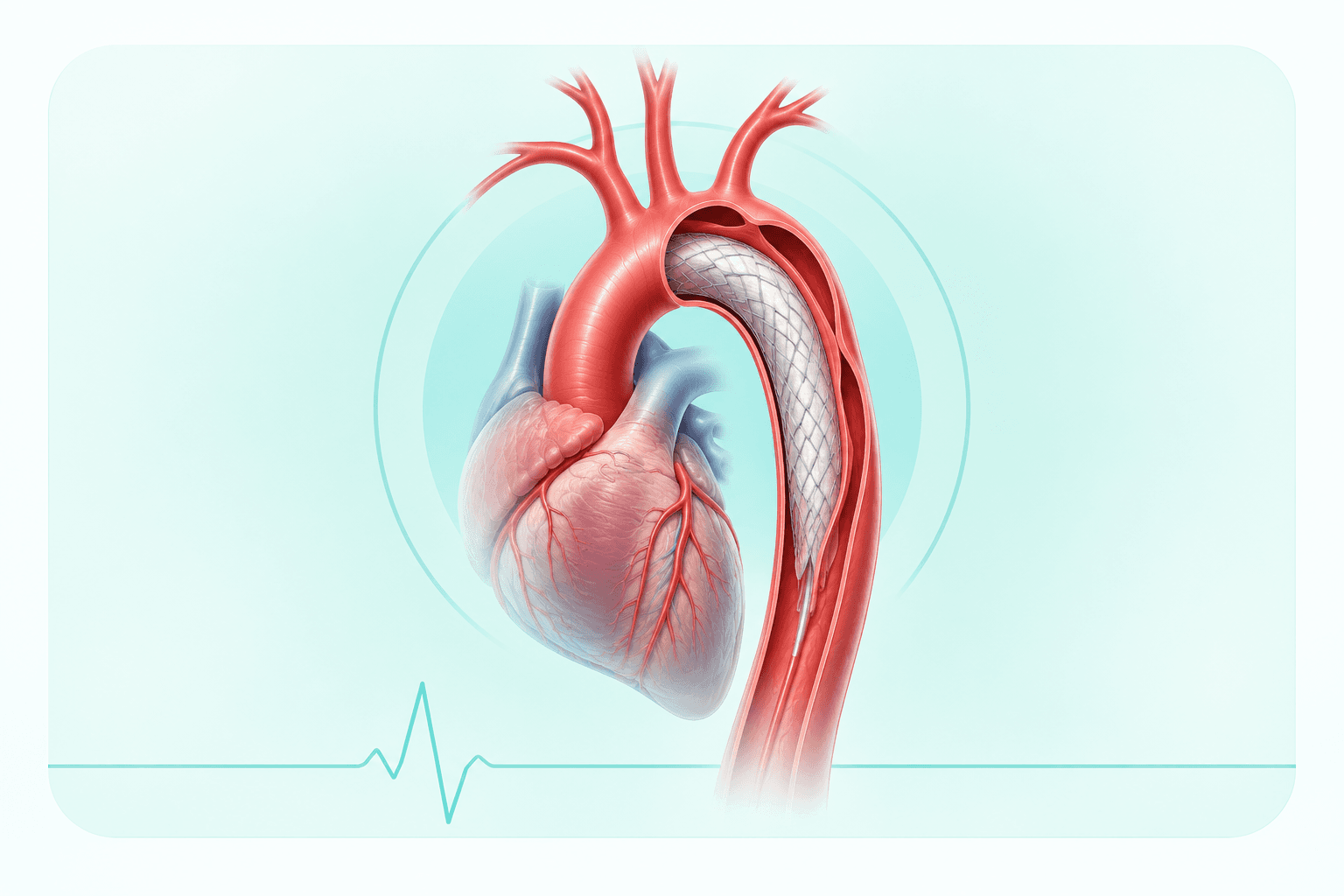
As one of the few surgeons in India with specialised training in complex aortic surgery, Dr. Idhrees treats aortic aneurysms, acute aortic dissections, and performs advanced procedures such as the frozen elephant trunk technique for arch repair.
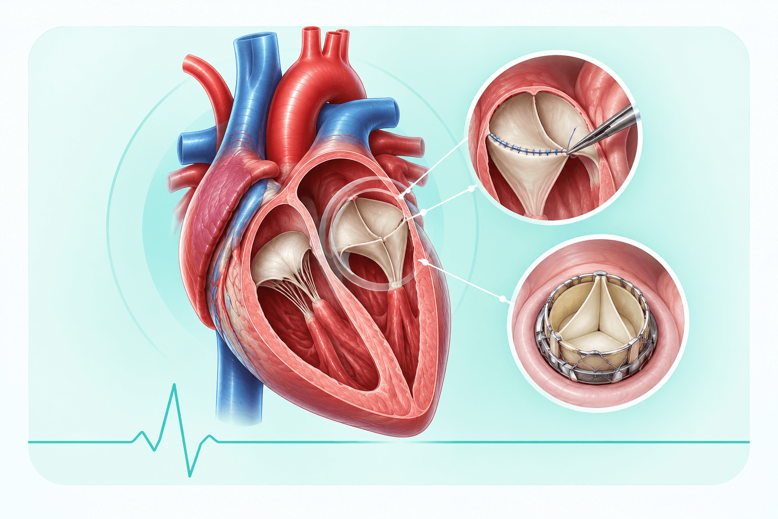
Expert surgical repair and replacement of damaged or diseased heart valves — including mitral, aortic, and tricuspid valve surgery — restoring normal heart function with a focus on long-term durability.
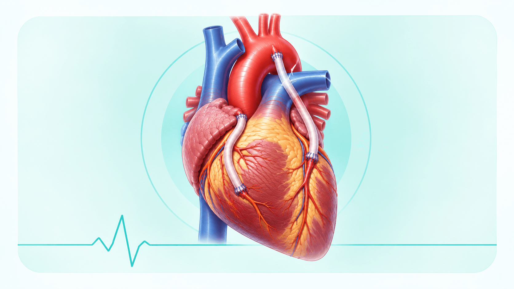
Dr. Idhrees performs advanced coronary artery bypass grafting (CABG) to restore healthy blood flow to the heart in patients with blocked coronary arteries, using both conventional and beating-heart (off-pump) techniques for faster recovery.